Pregnancy SmartSiteTM
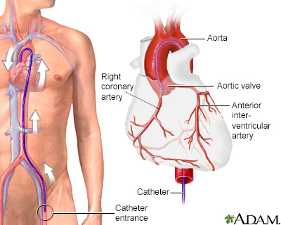
Angiography - aorta; Aortography; Abdominal aorta angiogram; Aortic arteriogram; Aneurysm - aortic arteriogram DefinitionAortic angiography is a procedure that uses a special dye and x-rays to see how blood flows through the aorta. The aorta is the large artery that carries blood from your heart to your belly, pelvis, and legs. Angiography uses x-rays and a special dye to see the insides of the arteries. Arteries are blood vessels that carry blood away from the heart. How the Test is PerformedThis test is done at a hospital or specialized procedure unit. Before the test starts, you will be given a mild sedative to help you relax.
After the x-rays or treatments are finished, the catheter is removed. Pressure is applied to the puncture site for 20 to 45 minutes to stop the bleeding. After that time, the area is checked and a tight bandage is applied. The leg is most often kept straight for another 6 hours after the procedure. You should avoid strenuous activity, such as heavy lifting, for 24 to 48 hours. How to Prepare for the TestYou may be asked not to eat or drink anything for 6 to 8 hours before the test. You will wear a hospital gown and sign a consent form for the procedure. Remove jewelry from the area being studied. Tell your health care provider:
You will be awake during the test. You may feel a sting as the numbing medicine is given and some pressure as the catheter is inserted. You may feel a warm flushing when the contrast dye flows through the catheter. This is normal and most often goes away in a few seconds. You may have some discomfort from lying on the hospital table and staying still for a long time. In most cases, you can resume normal activity the day after the procedure. Why the Test is PerformedYour provider may ask for this test if there are signs or symptoms of a problem with the aorta or its branches, including:
What Abnormal Results MeanAbnormal results may be due to:
RisksRisks of aortic angiography include:
ConsiderationsThis procedure may be done with left heart catheterization to look for coronary artery disease. Aortic angiography has been mostly replaced by computed tomography (CT) angiography or magnetic resonance (MR) angiography. ReferencesBraverman AC, Schemerhorn M. Diseases of the aorta. In: Libby P, Bonow RO, Mann DL, Tomaselli GF, Bhatt DL, Solomon SD, eds. Braunwald's Heart Disease: A Textbook of Cardiovascular Medicine. 12th ed. Philadelphia, PA: Elsevier; 2022:chap 42. Reekers JA. Angiography: principles, techniques and complications. In: Adam A, Dixon AK, Gillard JH, Schaefer-Prokop CM, eds. Grainger & Allison's Diagnostic Radiology. 7th ed. Philadelphia, PA: Elsevier; 2021:chap 78. van den Bosch H, Westenberg JJM, de Roos A. Cardiovascular magnetic resonance angiography: carotids, aorta, and peripheral vessels. In: Manning WJ, Pennell DJ, eds. Cardiovascular Magnetic Resonance. 3rd ed. Philadelphia, PA: Elsevier; 2019:chap 44. | |
| |
Review Date: 10/23/2024 Reviewed By: Deepak Sudheendra, MD, MHCI, RPVI, FSIR, Founder and CEO, 360 Vascular Institute, with an expertise in Vascular Interventional Radiology & Surgical Critical Care, Columbus, OH. Review provided by VeriMed Healthcare Network. Also reviewed by David C. Dugdale, MD, Medical Director, Brenda Conaway, Editorial Director, and the A.D.A.M. Editorial team. The information provided herein should not be used during any medical emergency or for the diagnosis or treatment of any medical condition. A licensed medical professional should be consulted for diagnosis and treatment of any and all medical conditions. Links to other sites are provided for information only -- they do not constitute endorsements of those other sites. No warranty of any kind, either expressed or implied, is made as to the accuracy, reliability, timeliness, or correctness of any translations made by a third-party service of the information provided herein into any other language. © 1997- A.D.A.M., a business unit of Ebix, Inc. Any duplication or distribution of the information contained herein is strictly prohibited. | |

 Cardiac arteriogra
