Pregnancy SmartSiteTM
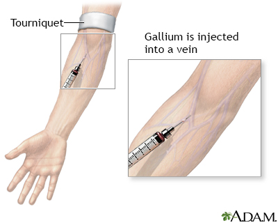
Gallium 67 lung scan; Lung scan; Gallium scan - lung; Scan - lung DefinitionLung gallium scan is a type of nuclear scan that uses radioactive gallium to identify inflammation in the lungs. How the Test is PerformedGallium is injected into a vein. The scan will be taken 6 to 24 hours after the gallium is injected. (Test timing depends on whether your condition is acute or chronic.) During the test, you lie on a table that moves underneath a scanner called a gamma camera. The camera detects the radiation produced by the gallium. Images are displayed on a computer screen. During the scan, it is important that you keep still to get a clear image. The technician can help make you comfortable before the scan begins. The test takes about 30 to 60 minutes. How to Prepare for the TestSeveral hours to 1 day before the scan, you will get an injection of gallium at the place where the testing will be done. Just before the scan, remove jewelry, dentures, or other metal objects that can affect the scan. Take off the clothing on the upper half of your body and put on a hospital gown. How the Test will FeelThe injection of gallium will sting, and the puncture site may hurt for several hours or days when touched. The scan is painless, but you must stay still. This may cause discomfort for some people. Why the Test is PerformedThis test is usually done when you have signs of inflammation in the lungs. This is most often due to sarcoidosis or a certain type of pneumonia. It is not performed very often in recent years. Normal ResultsThe lungs should appear of normal size and texture, and should take up very little gallium. What Abnormal Results MeanIf a large amount of gallium is seen in the lungs, it may mean any of the following problems:
RisksThere is some risk to children or unborn babies. Because a pregnant or nursing woman may pass on radiation, special precautions need to be taken. For women who are not pregnant or nursing and for men, there is very little risk from the radiation in gallium, because the amount is very small. There are increased risks if you are exposed to radiation (such as x-rays and scans) many times. Discuss any concerns you have about radiation with your health care provider who recommends the test. ConsiderationsUsually your provider will recommend this scan based on the results of a chest x-ray. Small defects may not be visible on the scan. For this reason, this test is not often done anymore. ReferencesHarisinghani MG, Chen JW, Weissleder R. Chest imaging. In: Harisinghani MG, Chen JW, Weissleder R, eds. Primer of Diagnostic Imaging. 6th ed. Philadelphia, PA: Elsevier; 2019:chap 1. Jokerst CE, Gotway MB. Thoracic radiology: noninvasive diagnostic imaging. In: Broaddus VC, Ernst JD, King TE, et al, eds. Murray and Nadel's Textbook of Respiratory Medicine. 7th ed. Philadelphia, PA: Elsevier; 2022:chap 20. | |
| |
Review Date: 8/19/2024 Reviewed By: Allen J. Blaivas, DO, Division of Pulmonary, Critical Care, and Sleep Medicine, VA New Jersey Health Care System, Clinical Assistant Professor, Rutgers New Jersey Medical School, East Orange, NJ. Review provided by VeriMed Healthcare Network. Also reviewed by David C. Dugdale, MD, Medical Director, Brenda Conaway, Editorial Director, and the A.D.A.M. Editorial team. The information provided herein should not be used during any medical emergency or for the diagnosis or treatment of any medical condition. A licensed medical professional should be consulted for diagnosis and treatment of any and all medical conditions. Links to other sites are provided for information only -- they do not constitute endorsements of those other sites. No warranty of any kind, either expressed or implied, is made as to the accuracy, reliability, timeliness, or correctness of any translations made by a third-party service of the information provided herein into any other language. © 1997- A.D.A.M., a business unit of Ebix, Inc. Any duplication or distribution of the information contained herein is strictly prohibited. | |

 Gallium injection
